Zika Virus and Blood Transfusions

 Zika and Blood Transfusions: The Current Landscape
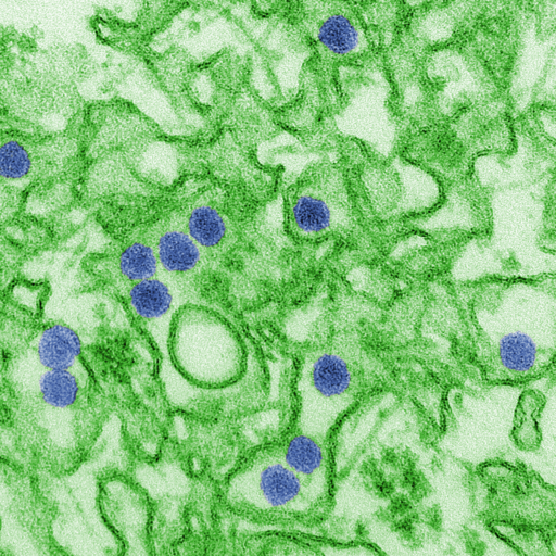
Scanning Electron Microscope image of Zika

Zika virus, initially identified in the Zika forest of Uganda in 1947, emerged as a significant global health concern during the outbreak that began in Brazil in 2015. Transmitted primarily through the bite of an infected Aedes species mosquito, Zika garnered international attention due to its association with microcephaly in newborns and other severe birth defects. Additionally, concerns were raised about the potential for Zika virus transmission through blood transfusions, creating an urgency in the blood banking community to implement safety measures.

Zika Transmission through Blood Transfusions

While the majority of Zika virus cases result from mosquito bites, several instances of transmission through blood transfusions were reported during the major outbreaks. The virus can survive and remain infectious in blood products, making the transfusion of contaminated blood a possible transmission route. Furthermore, a significant number of infected individuals are asymptomatic, which makes it harder to identify and defer potentially infectious donors based solely on clinical symptoms.

Blood Screening for Zika

Given the potential risks associated with Zika virus transmission through transfusions, many countries, particularly those with reported Zika cases, initiated blood screening procedures to ensure the safety of the blood supply.

In the United States, the Food and Drug Administration (FDA) issued guidance in 2016 recommending universal testing of all donated Whole Blood and blood components for Zika virus in the states and territories with active transmission. By 2018, this guidance expanded to include universal testing across all states and territories, regardless of the presence of active Zika cases. This approach utilized nucleic acid testing (NAT) to detect the presence of the virus in donated blood.

Current Testing Guidelines

Over time, as the number of Zika cases declined and a better understanding of the virus and its transmission patterns emerged, the FDA updated its guidelines. In 2019, the FDA revised its recommendations, allowing blood centers to test pooled samples rather than individual donations. This shift was based on risk-assessment models showing a significant decrease in the prevalence of Zika virus infection among blood donors in the U.S.

Internationally, blood screening protocols vary based on the prevalence of the virus, available resources, and the assessed risk of transfusion-transmitted Zika virus. Many countries with no reported cases or those that have never experienced local transmission might not routinely screen blood donations for Zika.

Implications for Blood Safety

The introduction and continuous update of Zika screening protocols signify the nimbleness required in transfusion medicine. In the face of emerging infectious threats, the blood banking community must be prepared to quickly assess risks and implement appropriate safety measures.

Zika's emergence reinforced the importance of proactive measures, research, and international collaboration to ensure the safety of the blood supply. While the immediate crisis associated with the Zika virus has subsided, the lessons learned continue to shape policies and preparedness strategies for future threats.

The Effects of Zika Virus

While many people infected with Zika virus remain asymptomatic or experience only mild symptoms, the real concern lies in the complications linked to the virus:

  1. Birth Defects: One of the most alarming complications associated with Zika is its ability to cause congenital disabilities when pregnant women contract the virus. Microcephaly, where a baby's head is much smaller than expected, is the most recognized of these birth defects. Infants with microcephaly often have underdeveloped brains, leading to long-term developmental challenges and sometimes even death.

  2. Guillain-Barré Syndrome (GBS): Zika has been associated with GBS, a rare neurological disorder where a person's immune system attacks their nerves. GBS can result in muscle weakness and, in severe cases, paralysis. Though most people recover from GBS, some might experience long-term effects, and in rare cases, it can be fatal.

  3. Other Neurological Complications: Apart from GBS, Zika has been linked to other neurological conditions such as meningoencephalitis and myelitis.

Therapeutic Phlebotomy

Therapeutic Phlebotomy: Controlled Blood Removal as Treatment

Therapeutic phlebotomy, often simply termed as "bloodletting," has been practiced for centuries, dating back to ancient civilizations like the Egyptians and Greeks. They believed that removing blood from the body could rid it of evil spirits or balance out the four humors: blood, phlegm, black bile, and yellow bile. While modern medicine has debunked these ancient theories, controlled blood removal remains a medically approved treatment for specific conditions. One such condition is polycythemia vera, but there are several others where therapeutic phlebotomy has proven beneficial.

 The Procedure

Undergoing therapeutic phlebotomy is similar to giving blood at a blood donation center. The patient is comfortably seated, and a large needle is inserted into a vein, typically in the arm. Blood is then drawn into a bag. The entire process can last anywhere from 30 minutes to an hour, depending on the volume of blood being removed.

It's essential to drink plenty of fluids before and after the procedure to help replace the lost volume and prevent feelings of dizziness or lightheadedness.

Patient Experiences

Patients undergoing therapeutic phlebotomy often report a range of experiences. Some feel revitalized, while others might feel tired immediately after the procedure. Temporary dizziness or lightheadedness is not uncommon. For those with conditions like hemochromatosis, they often report feeling 'lighter' and less fatigued as excess iron is removed from their system. Over time, with regular phlebotomies, symptoms related to their underlying condition, such as joint pain in hemochromatosis or itching in polycythemia vera, often diminish.

Risks and Side Effects

As with any medical procedure, there are potential risks involved. These can include:

  • Bruising at the needle site
  • Infection, although this is rare if proper aseptic techniques are used
  • Feeling faint or dizzy
  • Hematoma (a collection of blood outside of blood vessels)

It's also worth noting that over time, veins can become scarred or harder to access. In such cases, a phlebotomist or healthcare provider might choose a different site or vein for blood removal.

Polycythemia Vera (PV)

Polycythemia vera is a rare type of blood cancer where the bone marrow produces too many red blood cells. This overproduction thickens the blood, slowing its flow and increasing the risk of clots. These clots can lead to severe complications such as heart attacks or strokes. By regularly removing a specific volume of blood, therapeutic phlebotomy reduces the number of circulating red blood cells. This procedure brings the hematocrit closer to normal levels, reducing the associated risks. Patients might require frequent phlebotomies initially, with the frequency tapering off as the condition stabilizes.

Hemochromatosis

Hemochromatosis is a genetic disorder causing the body to absorb too much iron from food. Over time, this excess iron accumulates in organs, particularly the liver, heart, and pancreas, leading to complications such as liver disease, heart problems, or diabetes. Through therapeutic phlebotomy, blood is removed regularly to decrease the body's iron levels. Blood may be removed weekly until iron levels normalize, and maintenance sessions might follow every 2-4 months.

Porphyria Cutanea Tarda (PCT)

PCT, the most common type of porphyria, arises from a deficiency of the enzyme uroporphyrinogen decarboxylase. Patients exhibit symptoms like blisters on sun-exposed skin, excessive hair growth, and liver abnormalities. Elevated liver iron stores often accompany PCT, and iron plays a role in its manifestation. Therapeutic phlebotomy serves to reduce these iron stores, subsequently decreasing porphyrin levels in the liver and mitigating symptoms.

Secondary Polycythemia

Beyond the primary polycythemia vera, there's secondary polycythemia, where red blood cell overproduction results from external factors, such as chronic hypoxia or tumors secreting erythropoietin. Therapeutic phlebotomy can address the excessive red blood cells, offering symptomatic relief and reducing potential complications.

Patient Monitoring and Individualized Care

For those undergoing therapeutic phlebotomy, regular monitoring is crucial. It ensures that optimal hematocrit or iron levels are maintained and complications are avoided. Factors like the patient's age, overall health, and the underlying condition dictate the frequency and volume of blood removal. Regular blood tests and clinical assessments guide adjustments in the phlebotomy regimen, ensuring each patient receives care tailored to their unique needs.

Polycythemia Vera


What is Polycythemia Vera?


Polycythemia vera (PV) is an uncommon blood cancer characterized as a myeloproliferative neoplasm. What this means is that the bone marrow has uncontrolled and abnormal cellular proliferation or growth. This causes the overabundance of cells being created to push out of the bone marrow crowding the vascular system with excess cells. In the case of Polycythemia vera there is an increase in Red Blood Cells (erythrocytes) but also White Blood Cells (leukocytes), as well as platelets (thrombocytes). It is most notable that unlike most other blood cancers (such as leukemias) Red Blood Cells are markedly increase, which is a hallmark of the disease. This extreme increase in circulating Red Blood Cells accounts for most of the signs and symptoms seen with Polycythemia vera. Polycythemia in itself means "many cells in the blood". Erythrocytosis is another term for Polycythemia. 

In general PV is seen and diagnosed more often in men than women. It is rarely seen in those under 40 years of age and becomes more prevalent as aging continues. In a given year, 1 in 36000 men will be diagnosed with PV, while just 1 in 77,000 women will be diagnosed. Roughly 1 out of 4500 people at any given time will be affected by the disease.

 PV is generally not an inheritable disorder and would not be expected to be passed down through generations. Like most cancers it's inception lies in a somatic genetic mutation that causes the bone marrow to overproduce Red Blood Cells. Being a somatic mutation, this means that the mutation occurred post-conception and was not a genetic mutation present at conception or birth, but as a result of a gene mutating during normal cell replication.

The vast majority of patients with PV have a mutation in the Janus Kinase 2 gene, also known as JAK2. JAK2 is an important protein involved in cell signaling. It is involved with regulating cell growth and replication as well as regulating cell production with the confines of the bone marrow. A mutation in this gene is like essentially losing the ability to say STOP! Without the ability of JAK2 to regulate cell production, the cells grow and divide and create more and more cells without cease. Cells continue to be made even in absence of erythropoietin, the hormone responsible for signaling that more RBCs should be produced. 

Care must be taken not to diagnose the etiology of polycythemia (or erythrocytosis) incorrectly. Polycythemia refers to a general condition in which the overall Red Blood Cell mass is increased within the patients circulation. Polycythemia vera refers to the malignant overproduction of cells within the bone marrow, a cancerous process. Vera in Latin translates to "True". So Polycythemia vera is a true polycythemia caused by a malignant cancerous process and could also be referred to as PRIMARY Polycythemia. Secondary polycythemia may occur, causing increased Red Blood Cell and Hemoglobin counts, but is not related to a cancerous process. 

Non-cancerous polycythemia/erythrocytosis can be caused by other factors that cause erythropoietin to be made, which is a hormone that tells the bone marrow to stimulate bone marrow. Conditions such as sleep apnea or COPD in which oxygen enters the blood at a lower rate, could trigger the body to produce more Red Blood Cells. Smoking, Alcohol use, diuretics can sometimes cause an Apparent Polycythemia. Chronic dehydration can also cause an apparent polycythemia, as the liquid portion of blood (plasma) is reduced. 

Signs and Symptoms


Many patients with Polycythemia vera may not know they have it. It is generally a slow and eventual process.  It may not be until they have routine blood work done, such as a CBC (Complete Blood Count), that their doctor is tipped off to something being wrong. The CBC of a patient with PV may show increased absolute Red Blood Cell count, increased Hemoglobin, as well as an increase in hematocrit percentage. Hemoglobin is major protein within a Red Blood cell that not only gives Red Blood cells their red hue, but it is also responsible for carrying oxygen throughout the body and to the tissues. It's no wonder that if the amount of Red Blood Cells increase, hemoglobin will generally follow and increase as well. The Hematocrit is a percentage of Red Blood Cells related to the total amount of blood volume. The remaining portion of blood volume that isn't Red Blood Cells, is mostly plasma. For some with PV, they can see a slight but generally not malignant increase in White Blood Cells as well as platelets. 

Normal RBC counts are roughly 4.5 to 6.0×10^12/L for men, and 4.0 to 5×10^12/L for women. Normal hemoglobin ranges 14 to 17 grams per deciliter(dL) for men and 12 to 15 grams per deciliter for women. Hematocrits range from 40-50% in men and 35-45% in women. Counts above this range, coupled with other symptoms could lead a doctor to suspect Polycythemia vera. Ranges will differ slightly based on location due to slight differences in instrumentation and patient population. 

Normal Blood Counts per MayoClinic laboratories


Polycythemia vera is dangerous however, because with an increase of Red Blood Cells beyond what is normal, the blood starts to become thicker and less viscous. This makes circulating the blood throughout the body much more difficult and can lead to dangerous situations such as blood clots and strokes. The situation in which the thickening, or an increase of viscosity of the blood, is called hyperviscosity syndrome. It is not specific to PV, and patient's with other hematologic malignancies such as Waldenstroms macroglobulinemia, leukemias, Multiple Myeloma, etc. Patient's with PV that start to experience hyperviscosity syndrome can expect to see most non-specific symptoms such as 
  • Headache
  • Nausea
  • Vision Changes and retinopathy
  • Dizziness/Vertigo
  • Seizures and potentially even coma. 
  • Dyspnea (Shortness of breath)
  • Fatigue/Weakness
  • Stroke
  • Night Sweats
  • Bruising / Extended Bleeding
  • Weight Loss

Nearly one fifth of all patients with PV are found to have clots within their circulatory system. A large majority of clots can cause and/or be related to stroke or TIAs (Transient Ischemic Attacks aka Mini-stroke).

Increased blood viscosity in PV is related to decreased blood flow (ischemia), especially to the brain which leads to an increased risk of thrombotic (clotting) events. Similarly, clots and hyperviscous blood can cause organ damage throughout the body, as well as heart attacks. 

Other symptoms include

Erythromelalgia with blotchy, discolored, red skin. This is due to the increase of red blood cells in the body and the slower circulation of the cells. The skin may be warm to the touch as well. A burning pain sensation can also occur as parts of the vasculature in the extremities can get periodically blocked due to the viscous blood. 

Itchy skin, especially after a warm bath/shower -- nearly half of all patients with PV will have itchy skin. It can be the first symptom to show before the disease progresses. It is thought that this is caused by an abnormal function of histamine release as well as cytokine release. (cytokines are cell signaling molecule)

Gout -- gout is a painful type of arthritis caused by an excess buildup of uric acid in the body. Uric acid crystals become deposited in areas of a patient's joints, usually causing immense pain. The big toe is the most common joint to experience this. In general gouty arthritis this is usually caused by the bodies failure to remove uric acid from circulation such as seen with a low functioning kidney. It can also be due to excessive intake through foods, etc. In the case of PV, it is related to the high turnover of Red Blood Cells causing excess UA to be produced,  and potentially the decreased excretion of Uric Acid due to organ damage related to PV. Increased Uric Acid and gout is also related to the myelofibrosis that can sometimes be seen in PV patients.

Splenomegaly -- an enlarged spleen. One of the spleen's important functions is filtering out and removing old or damaged Red Blood Cells. In PV, since there are so many Red Blood Cells, there is an marked increase in the amount of old/damaged Red Blood Cells that the spleen has to process. This causes the spleen to enlarge as it becomes overworked and the cells build up. If it becomes too large, patient's can feel discomfort in that area.  

Myelofibrosis --- a severe scarring of the bone marrow that can occur as part of the PV disease progression. Myelofibrosis is not unique to PV and can happen under many other conditions. Over time, after consistently overproducing cells for extended periods of time, the bone marrow can become so overrun by scar tissue from the damaging effects of over proliferation, that it is no longer able to effectively make cells. This results in a broad DECREASE in cells across the board. One could expect to see decreased Red Blood Cell counts at this point (resulting in anemia), as well as a decrease in White Blood Cell counts and Platelets. Not all patients will progress to Myelofibrosis but nearly 10% may. Myelofibrosis also increases the risk of having gout symptoms as seen above. 

Diagnosis

Many patients with Polycythemia vera may not know they have the disease for some time. Initial symptoms may come and go, or be vague enough to not pinpoint to any one problem until the disease progresses. 

Some many initially present with a headache, fever, itchy/warm/red skin, dizziness, etc. Some patients may have an obvious abdominal bulge due to splenomegaly. 

As part of any workup, the doctor will almost always order a CBC (Complete Blood Count). Since Polycythemia vera's hallmark is the presence of an increased Red Blood Cell mass, a CBC would be an excellent tool in aiding in the diagnosis of PV. For men, a Hemoglobin concentration of greater than 16.5 g/dL, a hematocrit above 50%, and an increased total Red Blood Cell count would be highly indicative for disease. For women, normal hemoglobin and hematocrit levels are slightly lower than men so roughly 16g/dL and 48% hematocrit could potentially raise alarm.

 If the doctor can rule out secondary reasons for these being increased, such as dehydration, these numbers would be a cause for your doctor to potentially follow closer to a Polycythemia vera diagnosis. Results far above the previously mentioned numbers would likely aid in diagnosis. Numbers that high but borderline normal, may need more probing. Presenting with any of the symptoms listed here would likely aid in diagnosis. Many instances of increased Hemoglobin/Red Cells/Hematocrit are not due to Polycythemia vera, but to secondary processes. 

Your doctor may want to test for mutations within the Janus Kinase 2 (JAK2) gene. As previously mentioned, this gene is responsible for creating a protein that is involved in cell signaling. It tells the bone marrow to "start" or "stop" the creation of new blood cells. A very common mutation "JAK2(V617F)" is found in the overwhelming majority of patients with Polycythemia vera. A laboratory can test your blood and search for this mutation within the gene. It is also possible for the laboratories to search for other metalations within the gene if testing comes up negative for the JAK2(V617F) mutation allele. 

Erythropoietin -- a hormone that signals for the body to create more red blood cells. Patients with PV will generally have very low levels of EPO, given that EPO works off a feedback loop, if red cells and tissue oxygenation are abundant there's no need for more EPO to be created. Patients with high RBC/Hemoglobin(Hgb)/Hematocrit(Hct) counts suffering from dehydration, COPD, sleep apnea, etc., will not have these severely low levels. Your doctor may use JAK2 mutation in conjunction with EPO testing to be sure. 

Bone Marrow Biopsy -- your doctor may recommend a bone marrow biopsy and/or bone marrow aspirate. A needle is inserted in the bone, generally through the area of the Iliac Crest on the hip. This is the most common and safest site for biopsy due to its location away from major organs and blood vessels. The aspirate portion of the procedure involves removing some of the fluid portion of the marrow itself for examination. Slides will generally be made from this to be viewed microscopically. During the same procedure, a piece of solid bone marrow may be removed for analysis. The end results would generally show a bone marrow with far too many Red Blood Cells being created at a given time.

Treatment 

Treatment of Polycythemia vera will depend on certain things like overall health and age, severity of disease and likelihood of progression, and other risk factors. 

Those of younger age, typically under 60 with no history of clotting disorders or events are generally considered lower risk than those of more advanced age and/or those with previous clotting events. 

Unfortunately there is no definitive cure for PV. There are a number of treatments available to lessen symptoms and improve outcomes. PV carries a fairly optimistic prognosis unlike some cancers and hematological malignancies. On average, most people can live twenty years past their diagnosis date if they remain committed to their health and treatment plan. The most common cause of death in PV patients is clotting, so it is essential to be on top of things post-diagnosis. Other complications include progressing disease which may transform into myelofibrosis, AML (Acute Myelogenous Leukemia), or MDS (Myelodysplastic Syndrome). The treatments available generally do not lessen the risk of progression towards AML or MDS. Roughly 10 percent of patient's with PV may experience transformation info AML or MDS. Patients who progress to AML or MDS will see shorted expectations and worse prognosis. 

Therapeutic Phlebotomy -- It is exactly as it sounds. When you go to the doctor and have labs drawn, a nurse or phlebotomist will perform a phlebotomy or "venipuncture" to acquire blood from the patient for testing. This is essentially the same idea. Therapeutic phlebotomy involves a venipuncture in which blood is removed from the patient in order to relieve many of the symptoms and potential future problems of having hyperviscous blood. 
 
Up to 500mL is removed during a single sitting (the same amount removed during blood donation). Lesser amounts may be taken depending on the patient's cardiovascular heath and age. Initially, phlebotomies will take place as often as possible (once a day, every other day) until the patient's hematocrit reaches a normal 45%. From there a plan will be devised and often patients will only need phlebotomies once every month or every other month. It is possible however that due to frequent phlebotomies, iron within the blood may decrease. This treatment alone can buy the patient a large amount of their life back. Many of the symptoms listed here will be alleviated by having normal blood levels. This does not prevent transformation into other cancers such as AML/MDS however. For low risk patients, this is one of the only treatments that may be necessary. No drugs or chemotherapies needed.


Aspirin therapy -- for low risk patients without a history of bleeding issues, low dose aspirin may be prescribed. Aspirin inhibits platelet function which allows for better management of clotting in PV patients who have a much higher risk of clotting. Platelets are fundamental in the clotting process, thus why this is beneficial. 

Antihistamines -- This can be beneficial in treating patients seeing an increase in itchiness, especially after warm water contact

Hydroxyurea -- Hydroxyurea (HU) or hydroxycarbamide is a drug taken by mouth that is generally prescribed to patients with higher risk factors. It is a myelosuppressive drug which means that it suppresses the bone marrow from producing cells. It does this by inhibiting the production of DNA within the cells, which in turns means the cells can no longer replicate and grow. This will eventually produce lower RBC counts as well as other cells as well. One would expect platelet counts to drop as well (since elevated platelet counts are often seen in PV). One caveat to HU is that there may be a link to hydroxyurea use and progression to leukemia. Most studies show this risk is very small or non-existent. Often times, symptomatic Sickle Cell Anemia patients will be on this drug as well. 

Ruxolitinib -- also known as Jakafi or Jakavi is a janus kinase inhibitor. It will selectively inhibit JAK1 and JAK2 enzymes from functioning which will assist in inhibiting those with JAK2 enzymes that have formed from mutated JAK2 genes. If you remember from earlier reading on the main page, this JAK2 mutation causes it to be stuck "on" and signals to the bone marrow to perpetually create cells. Ruxolitinib would help shut down this activity and potentially help the bone marrow cease the overproduction of cells. Generally this drug is given for patients who do not respond to Hydroxyurea or cannot receive Hydroxyurea due to allergy or other negative response. 

Ropeginterferon Alfa-2b-njft (BESREMi) -- A form of Interferon alfa. It interacts with receptors within the bone marrow to modulate cell signaling the promotes "anti-proliferative" effects as well as immune modulating and pro-apoptotic (cell death) events. This was recently approved by the FDA in 2021 as an official treatment.  


CAR-T Therapy

CAR-T Therapy: From Concept to Clinical Application

The Genesis of CAR-T CAR-T, which stands for Chimeric Antigen Receptor T-cell therapy, represents a groundbreaking evolution in cancer treatment. Rooted in immunotherapy principles, CAR-T pivots on the idea of harnessing the body's immune system, specifically the T-cells, and re-engineering them to become adept fighters against cancer cells.

A Closer Look at T-cells and CAR T-cells are white blood cells and stalwarts of the adaptive immune system. Their primary function is to identify foreign threats and mount an immune response. However, cancer cells, due to their genetic similarity to normal cells, often manage to evade this detection.

The Chimeric Antigen Receptor (CAR) is a synthetic, bioengineered receptor. When introduced into T-cells, CAR provides them the ability to recognize specific antigens, predominantly present on the surface of cancer cells. This equips the T-cells with a targeting system, enabling them to locate and destroy cancer cells with heightened precision.

Apheresis: Harvesting the Fighters The journey of CAR-T therapy begins with the extraction of the patient's T-cells, a process known as apheresis:

  • Mechanics of Apheresis: This procedure, while resembling standard blood donation, is more intricate. Blood is extracted and passed through a machine where white blood cells, specifically T-cells, are separated. The rest of the blood is then infused back into the patient.

  • Duration and Output: Depending on the patient's health, T-cell count, and specific therapeutic protocol, apheresis can span several hours. The objective is to secure an optimal number of T-cells for the subsequent genetic modification.

Transition to Specialized Laboratories Post-apheresis, the extracted T-cells are primed for their transformative journey:

  • Transport Dynamics: The viability of T-cells during transportation is paramount. Rigorous temperature controls, along with quick transportation methods, are employed to ensure the cells remain functional upon arrival at the labs.

  • T-cell Modification: Within these state-of-the-art labs, the T-cells undergo genetic engineering. Using viral vectors or other methodologies, the CAR is integrated into the T-cells, enabling them to target specific cancer antigens.

  • Culturing and Expansion: After successful genetic modification, these T-cells are cultured in specialized environments, promoting their growth and multiplication. This ensures a robust army of CAR-T cells for reinfusion into the patient.

Reintroduction and the Fight Within The genetically modified T-cells, now termed CAR-T cells, are ready for reintroduction:

  • Preparing the Battlefield: Patients might undergo a 'lymphodepleting' chemotherapy regimen before CAR-T cell infusion. This creates a more favorable internal environment, enhancing the efficacy and proliferation of infused CAR-T cells.

  • Reinfusion Dynamics: Administering the CAR-T cells is similar to a blood transfusion. Once inside, these engineered cells commence their mission, detecting, and decimating targeted cancer cells.

Navigating Challenges Despite its transformative potential, CAR-T therapy presents challenges:

  • Safety and Side Effects: The introduction of engineered cells and their aggressive action can lead to severe reactions. Cytokine release syndrome (CRS), characterized by a sudden surge in inflammatory cytokines, can be life-threatening in certain cases. Neurological side effects are also documented.

  • Complexity and Cost: The intricate multi-step process, from apheresis to genetic engineering in specialized labs, renders CAR-T therapy expensive. The complexity also demands an intricate logistical orchestration, from T-cell extraction to their reinfusion.

The Horizon of CAR-T As CAR-T therapy garners more clinical experience, research aims to enhance its safety profile, expand its applicability, and optimize its cost structure. There's ongoing exploration into targeting solid tumors, minimizing side effects, and introducing modulatory mechanisms to control CAR-T cell activity.

Moreover, advancements in biotechnology, genetic engineering, and immunology promise to refine the CAR-T process further, presenting hope for a broader range of malignancies and more accessible therapeutic options for patients globally.

By intertwining the principles of immunology, the precision of genetic engineering, and the clinical rigor of processes like apheresis, CAR-T therapy epitomizes the vanguard of personalized medicine in oncology.

Can I Get an Infection From A Blood Transfusion?

In the realm of medical treatments, blood transfusions stand out as both indispensable and miraculous. They rejuvenate lives, ensuring that surgeries proceed, that trauma victims survive, and that patients with blood disorders thrive. Yet, with this life-saving potential comes the inherent question: Is there a risk of infection?

Historical Context

The landscape of blood transfusions, particularly in the early days, had its pitfalls. In earlier decades, before robust testing mechanisms were implemented, blood recipients faced higher risks. HIV, for example, was transmitted via transfusions in the early days of the AIDS epidemic. However, over time, the blood donation and transfusion field underwent transformative changes to drastically minimize these risks.

Initial Defenses: The Donor Selection Process

Long before blood is drawn from a donor's arm, the process of ensuring its safety begins. Every donor undergoes a detailed interview and is required to answer a comprehensive questionnaire. This is meticulously designed to weed out potential risks based on travel history, medical background, and certain behavioral factors. This rigorous interview, while time-consuming, establishes the first line of defense against bloodborne pathogens.

Testing: The Scientific Vanguard against Infections

The post-donation phase sees each unit of blood subjected to an exhaustive battery of tests:

HIV: Sensitive assays detect both the virus's antibodies and its RNA, ensuring that even recent infections don't slip through.

Hepatitis B & C: Both antibody and nucleic acid tests are deployed, offering dual layers of detection.

Syphilis: Regarded as an age-old enemy, modern treponemal tests detect this infection with impressive accuracy.

Other Threats: Whether it's West Nile virus, HTLV, or emerging concerns like Zika, blood banks remain vigilant, incorporating new tests as threats evolve.

Window Periods: A Persistent Challenge

Infections have a 'window period'—a timeframe post-infection when tests might not detect the pathogen. As testing becomes more advanced, these windows shrink, but they still pose a challenge that blood banks grapple with. The emphasis is on reducing this window as much as possible.

From Microbes to Prions: Broadening the Horizon

While viruses and bacteria are primary concerns, other potential threats, like prions (which cause conditions like Creutzfeldt-Jakob Disease), demand attention. The insidious nature of prions, their resistance to conventional sterilization techniques, and their long incubation period pose unique challenges, prompting continued research and surveillance.

Safety in Numbers: Quantifying the Risk

Statistics offer a clearer perspective:

  • For HIV, the risk stands at roughly 1 in 1.5 million.
  • Hepatitis B: About 1 in 280,000.
  • Hepatitis C: Approximately 1 in 1 million.

It's crucial to understand that these figures, as low as they are, represent a worst-case scenario. Real-world risks are often even lower, thanks to multiple overlapping safety measures.

Global Collaboration: A United Front

Blood safety isn't an isolated endeavor. Blood banks, researchers, and policymakers worldwide collaborate, sharing data, strategies, and insights. This global network ensures that emerging threats are rapidly identified, and best practices are universally adopted.

Continuous Training: The Human Element

Behind every machine and every test are dedicated professionals. Their training isn't static. As technology evolves and new threats emerge, continuous education ensures that these professionals remain at the forefront of safety.

Ethical Considerations and Transparency

Blood banks prioritize not just physical safety but also ethical considerations. Donors are informed about the tests their blood undergoes and any resultant findings. This transparency fortifies the bond of trust between donors and blood banks.

Blood Products and Derivatives: Extended Safety

Beyond whole blood, various blood products (plasma, platelets, cryoprecipitate) are used in medicine. Each has its unique processing and testing protocols, but the emphasis on safety remains paramount across all products.

The Relentless Pursuit of Safety

In the vast medical tapestry, blood transfusions remain a beacon of both hope and safety. The multi-tiered safety nets, combined with unwavering dedication from professionals in the field, ensure that risks are minimized. While perfection might remain an aspirational goal, the blood transfusion community relentlessly marches towards it, ensuring that each unit transfused not only saves a life but also stands as a testament to rigorous safety and quality.