Does a Blood Transfusion Change Your DNA?

DNA and Blood?
The idea of a blood transfusion has fascinated the human psyche for years. With its potential to save lives, it also brings along questions, one of which is: Can a blood transfusion change your DNA? To address this query, we need to understand the intricate details of blood components, especially red blood cells (RBCs) and DNA.

DNA and Genetic Information: At the heart of every cell in our body lies a nucleus (except mature red blood cells, which we'll delve into shortly). Within this nucleus is deoxyribonucleic acid (DNA), the molecule that contains our genetic code. This code acts as an instruction manual for our body – from determining our hair color and height to more complex processes like metabolism.

Blood Transfusions and Their Components: Blood transfusions aren't just about transferring red blood cells. Blood comprises several components: red blood cells, white blood cells (WBCs), platelets, and plasma. When a person receives a blood transfusion, depending on the requirement, they could receive any of these components. The most common transfusion, however, is the red blood cell transfusion.

Mature Red Blood Cells and Their Lack of Nucleus: Red blood cells are unique. Unlike other cells in our body, mature RBCs do not have a nucleus. This absence is actually by design. RBCs are primarily responsible for transporting oxygen from the lungs to the rest of the body and carrying carbon dioxide from the body's tissues back to the lungs. To maximize space for the oxygen-binding molecule, hemoglobin, RBCs eject their nucleus as they mature. This means that mature RBCs do not possess DNA.

Given this, when a person receives a transfusion of mature red blood cells, they are not receiving any DNA from the donor in those RBCs.

Reticulocytes and Their Genetic Material: While mature RBCs lack a nucleus, their precursors, called reticulocytes, do contain a nucleus. Reticulocytes are immature RBCs, and as they mature into functional RBCs, they eventually lose their nucleus. In a typical blood transfusion, the vast majority of cells are mature RBCs. However, a small number of reticulocytes might also be present. While these reticulocytes contain DNA, their presence is minuscule in comparison to the total blood volume.

White Blood Cells and DNA Transfer: Unlike RBCs, white blood cells (WBCs) do have nuclei and, therefore, contain DNA. However, transfusions are usually filtered to reduce the number of WBCs due to concerns about potential immune reactions. So, the number of WBCs (and thus the amount of donor DNA) in transfused blood is minimal.

Does Donor DNA Integrate into Recipient Cells? Even if small amounts of donor DNA enter the recipient's bloodstream through transfused WBCs or reticulocytes, this DNA does not integrate into the recipient's cells or genetic code. Our DNA remains stable and unchanged. The donor DNA present in WBCs or reticulocytes would eventually be degraded and cleared from the recipient's body.

Implications and Misconceptions: There have been misconceptions and myths surrounding blood transfusions, especially in popular culture. Stories of individuals acquiring memories or traits of their donors are scientifically unfounded. A blood transfusion does not alter a person's genetic makeup or bestow them with the characteristics or memories of the donor.

COVID-19 vaccine:  Conspiracy Theories floating around have also said that the vaccine can alter or change your DNA. Welllll no it doesn't. Nor does receiving blood products from a patient who received the coronavirus vaccine. And no... You can't request unvaccinated blood.

So, In essence, a blood transfusion does not change a recipient's DNA. While there may be trace amounts of donor DNA present in the transfused blood due to the minuscule number of reticulocytes or white blood cells, these do not integrate with the recipient's DNA. The primary purpose of a transfusion is to replenish the body's supply of essential blood components, especially red blood cells, to ensure the effective transport of oxygen throughout the body. Understanding the nuances of how transfusions work and their impact on the body is crucial in dispelling myths and emphasizing the life-saving potential of this medical procedure.

How Much Does a Blood Transfusion Cost?

If people donate blood for free why am I charged for a Blood Transfusion? 
Transfusions aren't free!

Well, it's complicated. But yes, there are charges involved. You are not charged for the blood itself, but for the process ensuring that the blood is safe, available, and transfusable. Blood transfusions can be costly, so it's crucial for a physician to determine if the transfusion is warranted.

Hospitals do NOT receive blood from blood suppliers for free. The blood supplier charges transfusion center blood banks for donation, manufacturing, and transportation. Red Blood Cells are typically purchased by a transfusion center from a Blood Donation Center at around $200-300, platelets can be roughly $600, and plasma units can be $40-60. The final cost to the patient may be up to 4 times this amount. These charges from the blood center and transfusion center aren't about taking advantage of the patient; there are many costs involved from donation to transfusion.

The blood center must hire employees to run blood drives and process the units. Every donation undergoes infectious disease laboratory testing. Each one must have its Blood Type identified. Component processing, tubings, bags, anticoagulants, shelf stability additives, bacterial testing, leukoreduction filters, and transporting blood to hospitals or other transfusion centers all come at a price.

On reaching the transfusion center, typically a hospital Blood Bank, the Blood Type must be confirmed via testing. The blood must be crossmatched to the patient by the Blood Bank. Sometimes, it may need to be irradiated, washed, or split (for pediatrics). Red Blood Cell units might also need antigen testing if a patient has an antibody towards common Red Cell Antigens.

Additionally, blood banks and transfusion services must meet stringent regulatory standards, which requires frequent training, audits, and quality controls. While ensuring the safety of the blood, it's also essential to account for the indirect costs, like utilities and administrative staff. The strict guidelines they follow are in place to maintain the highest level of safety and ensure the best care for patients.

Some units of collected blood might go unused due to positive tests for infectious diseases or simply because they expire. Given the limited shelf life of certain blood products, like platelets, there's an inherent cost associated with maintaining a larger inventory and managing unusable units.

Research is another essential facet of the blood transfusion world. Blood centers are constantly investing in ways to enhance safety, storage methods, and minimize reactions. While donors generously provide their blood, the system ensuring that this blood reaches those in need safely and efficiently isn't free.

In the end, a nurse will prep the patient for transfusion using a set that connects the bag of blood to a patient's IV. This set, including tubing, filter, and potentially a small bag of saline, also comes at a cost.

It's no small feat: a single transfusion of Packed Red Blood Cells can cost over $1000 after considering all the processes involved. So the next time you or someone you know needs a transfusion, remember the vast infrastructure that ensures each drop of blood is safe and ready when needed.

Zika Virus and Blood Transfusions

 Zika and Blood Transfusions: The Current Landscape

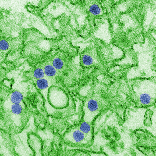
Scanning Electron Microscope image of Zika

Zika virus, initially identified in the Zika forest of Uganda in 1947, emerged as a significant global health concern during the outbreak that began in Brazil in 2015. Transmitted primarily through the bite of an infected Aedes species mosquito, Zika garnered international attention due to its association with microcephaly in newborns and other severe birth defects. Additionally, concerns were raised about the potential for Zika virus transmission through blood transfusions, creating an urgency in the blood banking community to implement safety measures.

Zika Transmission through Blood Transfusions

While the majority of Zika virus cases result from mosquito bites, several instances of transmission through blood transfusions were reported during the major outbreaks. The virus can survive and remain infectious in blood products, making the transfusion of contaminated blood a possible transmission route. Furthermore, a significant number of infected individuals are asymptomatic, which makes it harder to identify and defer potentially infectious donors based solely on clinical symptoms.

Blood Screening for Zika

Given the potential risks associated with Zika virus transmission through transfusions, many countries, particularly those with reported Zika cases, initiated blood screening procedures to ensure the safety of the blood supply.

In the United States, the Food and Drug Administration (FDA) issued guidance in 2016 recommending universal testing of all donated Whole Blood and blood components for Zika virus in the states and territories with active transmission. By 2018, this guidance expanded to include universal testing across all states and territories, regardless of the presence of active Zika cases. This approach utilized nucleic acid testing (NAT) to detect the presence of the virus in donated blood.

Current Testing Guidelines

Over time, as the number of Zika cases declined and a better understanding of the virus and its transmission patterns emerged, the FDA updated its guidelines. In 2019, the FDA revised its recommendations, allowing blood centers to test pooled samples rather than individual donations. This shift was based on risk-assessment models showing a significant decrease in the prevalence of Zika virus infection among blood donors in the U.S.

Internationally, blood screening protocols vary based on the prevalence of the virus, available resources, and the assessed risk of transfusion-transmitted Zika virus. Many countries with no reported cases or those that have never experienced local transmission might not routinely screen blood donations for Zika.

Implications for Blood Safety

The introduction and continuous update of Zika screening protocols signify the nimbleness required in transfusion medicine. In the face of emerging infectious threats, the blood banking community must be prepared to quickly assess risks and implement appropriate safety measures.

Zika's emergence reinforced the importance of proactive measures, research, and international collaboration to ensure the safety of the blood supply. While the immediate crisis associated with the Zika virus has subsided, the lessons learned continue to shape policies and preparedness strategies for future threats.

The Effects of Zika Virus

While many people infected with Zika virus remain asymptomatic or experience only mild symptoms, the real concern lies in the complications linked to the virus:

  1. Birth Defects: One of the most alarming complications associated with Zika is its ability to cause congenital disabilities when pregnant women contract the virus. Microcephaly, where a baby's head is much smaller than expected, is the most recognized of these birth defects. Infants with microcephaly often have underdeveloped brains, leading to long-term developmental challenges and sometimes even death.

  2. Guillain-Barré Syndrome (GBS): Zika has been associated with GBS, a rare neurological disorder where a person's immune system attacks their nerves. GBS can result in muscle weakness and, in severe cases, paralysis. Though most people recover from GBS, some might experience long-term effects, and in rare cases, it can be fatal.

  3. Other Neurological Complications: Apart from GBS, Zika has been linked to other neurological conditions such as meningoencephalitis and myelitis.

Therapeutic Phlebotomy

Therapeutic Phlebotomy: Controlled Blood Removal as Treatment

Therapeutic Phlebotomy
Therapeutic phlebotomy, often simply termed as "bloodletting," has been practiced for centuries, dating back to ancient civilizations like the Egyptians and Greeks. They believed that removing blood from the body could rid it of evil spirits or balance out the four humors: blood, phlegm, black bile, and yellow bile. While modern medicine has debunked these ancient theories, controlled blood removal remains a medically approved treatment for specific conditions. One such condition is polycythemia vera, but there are several others where therapeutic phlebotomy has proven beneficial.

 Therapeutic Phlebotomy Procedure

Undergoing therapeutic phlebotomy is similar to giving blood at a blood donation center. The patient is comfortably seated, and a large needle is inserted into a vein, typically in the arm. Blood is then drawn into a bag. The entire process can last anywhere from 30 minutes to an hour, depending on the volume of blood being removed.

It's essential to drink plenty of fluids before and after the procedure to help replace the lost volume and prevent feelings of dizziness or lightheadedness.

Patient Experiences

Patients undergoing therapeutic phlebotomy often report a range of experiences. Some feel revitalized, while others might feel tired immediately after the procedure. Temporary dizziness or lightheadedness is not uncommon. For those with conditions like hemochromatosis, they often report feeling 'lighter' and less fatigued as excess iron is removed from their system. Over time, with regular phlebotomies, symptoms related to their underlying condition, such as joint pain in hemochromatosis or itching in polycythemia vera, often diminish.

Risks and Side Effects of Therapeutic Phlebotomy

As with any medical procedure, there are potential risks involved. These can include:

  • Bruising at the needle site
  • Infection, although this is rare if proper aseptic techniques are used
  • Feeling faint or dizzy
  • Hematoma (a collection of blood outside of blood vessels)

It's also worth noting that over time, veins can become scarred or harder to access. In such cases, a phlebotomist or healthcare provider might choose a different site or vein for blood removal.

Polycythemia Vera (PV)

Polycythemia vera is a rare type of blood cancer where the bone marrow produces too many red blood cells. This overproduction thickens the blood, slowing its flow and increasing the risk of clots. These clots can lead to severe complications such as heart attacks or strokes. By regularly removing a specific volume of blood, therapeutic phlebotomy reduces the number of circulating red blood cells. This procedure brings the hematocrit closer to normal levels, reducing the associated risks. Patients might require frequent phlebotomies initially, with the frequency tapering off as the condition stabilizes.

Hemochromatosis

Hemochromatosis is a genetic disorder causing the body to absorb too much iron from food. Over time, this excess iron accumulates in organs, particularly the liver, heart, and pancreas, leading to complications such as liver disease, heart problems, or diabetes. Through therapeutic phlebotomy, blood is removed regularly to decrease the body's iron levels. Blood may be removed weekly until iron levels normalize, and maintenance sessions might follow every 2-4 months.

Porphyria Cutanea Tarda (PCT)

PCT, the most common type of porphyria, arises from a deficiency of the enzyme uroporphyrinogen decarboxylase. Patients exhibit symptoms like blisters on sun-exposed skin, excessive hair growth, and liver abnormalities. Elevated liver iron stores often accompany PCT, and iron plays a role in its manifestation. Therapeutic phlebotomy serves to reduce these iron stores, subsequently decreasing porphyrin levels in the liver and mitigating symptoms.

Secondary Polycythemia

Beyond the primary polycythemia vera, there's secondary polycythemia, where red blood cell overproduction results from external factors, such as chronic hypoxia or tumors secreting erythropoietin. Therapeutic phlebotomy can address the excessive red blood cells, offering symptomatic relief and reducing potential complications.

Patient Monitoring and Individualized Care

For those undergoing therapeutic phlebotomy, regular monitoring is crucial. It ensures that optimal hematocrit or iron levels are maintained and complications are avoided. Factors like the patient's age, overall health, and the underlying condition dictate the frequency and volume of blood removal. Regular blood tests and clinical assessments guide adjustments in the phlebotomy regimen, ensuring each patient receives care tailored to their unique needs.

Polycythemia Vera


What is Polycythemia Vera?

Definition and Characteristics: Polycythemia Vera (PV) is a rare type of blood cancer classified as a myeloproliferative neoplasm, which is characterized by the bone marrow's abnormal and uncontrolled cell growth. This results in an excessive production of blood cells, notably red blood cells (erythrocytes), but also white blood cells (leukocytes) and platelets (thrombocytes). A key feature of PV, distinguishing it from most other blood cancers, is the significant increase in red blood cells, a condition known as erythrocytosis or "many cells in the blood."

Prevalence and Demographics:

  • PV is more commonly diagnosed in men than in women.
  • It is rare in individuals under 40 years old and becomes more prevalent with age.
  • Annually, 1 in 36,000 men and 1 in 77,000 women are diagnosed with PV.
  • About 1 in 4,500 people are affected by PV at any given time.

Genetics and Mutation:

  • PV is generally not inherited and originates from a somatic genetic mutation post-conception.
  • Most patients with PV have a mutation in the Janus Kinase 2 (JAK2) gene, crucial for cell signaling and regulation within the bone marrow. This mutation leads to uncontrolled cell production.

Primary vs. Secondary Polycythemia:

  • Primary Polycythemia (Polycythemia Vera): A malignant overproduction of cells in the bone marrow due to a cancerous process.
  • Secondary Polycythemia: Caused by increased red blood cell production due to factors outside the bone marrow, such as low oxygen conditions or hormonal triggers, and not related to a cancerous process.

Non-Cancerous Causes: Conditions like sleep apnea, COPD, smoking, alcohol use, and the use of diuretics can lead to non-cancerous forms of polycythemia or erythrocytosis by stimulating the production of erythropoietin, a hormone that signals the bone marrow to produce more red blood cells.

Polycythemia Vera is a complex condition with specific genetic underpinnings and significant implications for blood cell production. Its diagnosis and treatment require careful differentiation from other forms of polycythemia, with a focus on managing the uncontrolled production of blood cells and the factors contributing to secondary forms of the condition.

Signs and Symptoms

Detecting Polycythemia Vera: Understanding Blood Work Indicators

Identification Challenges:

  • Many individuals with Polycythemia Vera (PV) may be unaware of their condition due to its gradual onset.
  • PV often comes to light during routine blood tests, like a Complete Blood Count (CBC), which can reveal anomalies hinting at the disease.

Key CBC Findings in PV:

  • Increased Red Blood Cell (RBC) Count: A significant indicator of PV, reflecting the overproduction of RBCs.
  • Elevated Hemoglobin Levels: Hemoglobin, the major protein in RBCs responsible for oxygen transport, also increases as RBC count rises.
  • Higher Hematocrit Percentage: Indicates a higher proportion of RBCs in blood volume. Hematocrit is the percentage of blood volume occupied by RBCs, with the remainder mostly being plasma.
  • Possible Increase in White Blood Cells and Platelets: Some individuals may also experience a slight, typically non-malignant, increase in these cell counts.

Normal Blood Count Ranges:

  • RBC Count: Approximately 4.5 to 6.0×10^12/L for men and 4.0 to 5×10^12/L for women.
  • Hemoglobin Levels: Typically 14 to 17 grams per deciliter (dL) for men and 12 to 15 g/dL for women.
  • Hematocrit Range: Between 40-50% for men and 35-45% for women.
  • It's important to note that normal ranges can slightly vary by location due to differences in lab equipment and population.

Clinical Implications:

  • Values above these normal ranges, along with other symptoms, may prompt a healthcare provider to suspect PV.
  • Such findings necessitate further investigation to confirm the diagnosis and understand the extent of the condition.

Normal Blood Counts per MayoClinic laboratories


Polycythemia vera is dangerous however, because with an increase of Red Blood Cells beyond what is normal, the blood starts to become thicker and less viscous. This makes circulating the blood throughout the body much more difficult and can lead to dangerous situations such as blood clots and strokes. The situation in which the thickening, or an increase of viscosity of the blood, is called hyperviscosity syndrome. It is not specific to PV, and patient's with other hematologic malignancies such as Waldenstroms macroglobulinemia, leukemias, Multiple Myeloma, etc. Patient's with PV that start to experience hyperviscosity syndrome can expect to see most non-specific symptoms such as 
  • Headache
  • Nausea
  • Vision Changes and retinopathy
  • Dizziness/Vertigo
  • Seizures and potentially even coma. 
  • Dyspnea (Shortness of breath)
  • Fatigue/Weakness
  • Stroke
  • Night Sweats
  • Bruising / Extended Bleeding
  • Weight Loss

Circulatory Clots:

  • About 20% of PV patients experience clots within their circulatory system, significantly increasing the risk of strokes and Transient Ischemic Attacks (TIAs).
  • The increased blood viscosity associated with PV leads to decreased blood flow and higher risk of thrombotic events, potentially causing organ damage and heart attacks.

Symptoms Overview:

  • Erythromelalgia: Characterized by blotchy, discolored, red skin, often warm to the touch and accompanied by a burning pain. This is due to increased red blood cells and slower circulation, causing periodic blockage in extremities.

  • Itchy Skin: Experienced by nearly half of all PV patients, often triggered after warm baths/showers. It may be an early symptom, believed to be caused by abnormal histamine and cytokine release.

  • Gout: A form of painful arthritis from uric acid buildup, commonly affecting the big toe. In PV, high RBC turnover and possible organ damage contribute to excessive uric acid production.

  • Splenomegaly: The spleen enlarges as it filters an excessive number of red blood cells, leading to discomfort or pain in the affected area.

  • Myelofibrosis: Severe scarring of the bone marrow seen in PV progression, leading to a broad decrease in cell production (anemia, reduced white cells, and platelets). Up to 10% of PV patients may develop myelofibrosis, further increasing gout risk.

Deeper Insights:

  • PV's impact on blood viscosity and cell proliferation poses significant risks, from increased clotting to organ strain.
  • Distinct symptoms like erythromelalgia and itchy skin after exposure to warmth highlight the body's stressed state due to excessive cell counts.
  • Conditions like gout and myelofibrosis underscore the systemic effects of PV, affecting everything from joint health to bone marrow function.
  • Understanding these symptoms and complications is crucial for managing PV and mitigating its impacts on patient health.

Recognizing and Diagnosing Polycythemia Vera (PV)

Early Detection Challenges:

  • Patients with Polycythemia Vera may not realize they have the disease due to intermittent or vague symptoms such as headaches, fever, itchy skin, or dizziness. Some may notice an abdominal bulge from splenomegaly.

Diagnostic Process:

  • A Complete Blood Count (CBC) is crucial for diagnosing PV, revealing increased Red Blood Cell (RBC) mass—a key indicator of the condition.
    • For Men: A hemoglobin concentration above 16.5 g/dL, hematocrit over 50%, and an increased RBC count suggest PV.
    • For Women: Hemoglobin around 16g/dL and hematocrit about 48% could signal the disease.
  • Ruling out secondary causes (e.g., dehydration) for increased blood counts is essential for a closer PV diagnosis. Abnormally high numbers, especially with symptoms, support the diagnosis, while borderline numbers require further investigation.

Advanced Testing for Confirmation:

  • JAK2 Mutation Testing: Testing for mutations in the Janus Kinase 2 (JAK2) gene is a critical step. The common mutation "JAK2(V617F)" is found in most PV patients. Laboratories can also search for other mutations if JAK2(V617F) is not present.

  • Erythropoietin (EPO) Levels: PV patients usually have very low EPO levels due to a feedback mechanism; abundant RBCs and oxygenation negate the need for more EPO production. High RBC/Hgb/Hct counts from other causes don't show this drastic EPO reduction. Testing EPO levels in conjunction with JAK2 mutations can confirm PV.

  • Bone Marrow Biopsy: A definitive diagnosis may require a bone marrow biopsy or aspirate, often taken from the iliac crest. This procedure examines the marrow directly, typically revealing excessive RBC production.

Key Points:

  • Initial symptoms of PV can be easily overlooked or misattributed, highlighting the importance of thorough medical evaluations for accurate diagnosis.
  • The CBC plays a foundational role in signaling potential PV, with further tests like JAK2 mutation analysis and EPO level measurement providing critical confirmation.
  • A bone marrow biopsy offers a direct look at marrow activity, confirming the excessive production of blood cells characteristic of PV.

Treatment of Polycythemia Vera

Understanding the Disease and Risks:

  • Polycythemia Vera is an incurable blood cancer with treatments available to manage symptoms and improve outcomes.
  • Life expectancy is generally optimistic, with many living over 20 years post-diagnosis, provided they adhere to their treatment plan.
  • The primary risk for PV patients is clotting, with disease progression to conditions like myelofibrosis, Acute Myelogenous Leukemia (AML), or Myelodysplastic Syndrome (MDS) as secondary concerns.

Risk-Based Treatment Approach:

  • Treatment varies based on factors like age, health, disease severity, and clotting history.
  • Patients under 60 without clotting issues are considered lower risk compared to older patients or those with previous clotting events.

Key Treatment Methods:

  • Therapeutic Phlebotomy:

    • Involves removing up to 500mL of blood to reduce blood viscosity and alleviate symptoms.
    • Frequency is initially high until hematocrit normalizes, then reduces based on individual needs.
  • Aspirin Therapy:

    • Low-dose aspirin may be prescribed to lower clotting risk by inhibiting platelet function, suitable for low-risk patients without bleeding issues.
  • Antihistamines:

    • Helps manage increased itchiness, especially following exposure to warm water.
  • Hydroxyurea (HU):

    • A myelosuppressive drug for higher-risk patients, inhibiting DNA production in cells to reduce RBC and platelet counts.
    • While concerns exist regarding its link to leukemia progression, evidence suggests minimal risk.
  • Ruxolitinib (Jakafi or Jakavi):

    • A JAK1 and JAK2 enzyme inhibitor, particularly useful for patients with JAK2 mutations leading to uncontrolled cell production.
    • Prescribed when Hydroxyurea is ineffective or unsuitable.
  • Ropeginterferon Alfa-2b-njft (BESREMi):

    • An FDA-approved treatment since 2021, BESREMi modulates cell signaling to have anti-proliferative, immune-modulating, and pro-apoptotic effects.


CAR-T Therapy

CAR-T Therapy: From Concept to Clinical Application

The Genesis of CAR-T 

CAR-T, which stands for Chimeric Antigen Receptor T-cell therapy, represents a groundbreaking evolution in cancer treatment. Rooted in immunotherapy principles, CAR-T pivots on the idea of harnessing the body's immune system, specifically the T-cells, and re-engineering them to become adept fighters against cancer cells.

A Closer Look at T-cells and CAR 

T-cells are white blood cells and stalwarts of the adaptive immune system. Their primary function is to identify foreign threats and mount an immune response. However, cancer cells, due to their genetic similarity to normal cells, often manage to evade this detection.

CAR-Engineered T-Cell Adoptive Transfer

The Chimeric Antigen Receptor (CAR) is a synthetic, bioengineered receptor. When introduced into T-cells, CAR provides them the ability to recognize specific antigens, predominantly present on the surface of cancer cells. This equips the T-cells with a targeting system, enabling them to locate and destroy cancer cells with heightened precision.

Apheresis: Harvesting the Fighters 

The journey of CAR-T therapy begins with the extraction of the patient's T-cells, a process known as apheresis:

  • Mechanics of Apheresis: This procedure, while resembling standard blood donation, is more intricate. Blood is extracted and passed through a machine where white blood cells, specifically T-cells, are separated. The rest of the blood is then infused back into the patient.

  • Duration and Output: Depending on the patient's health, T-cell count, and specific therapeutic protocol, apheresis can span several hours. The objective is to secure an optimal number of T-cells for the subsequent genetic modification.

Transition to Specialized Laboratories 

Post-apheresis, the extracted T-cells are primed for their transformative journey:

  • Transport Dynamics: The viability of T-cells during transportation is paramount. Rigorous temperature controls, along with quick transportation methods, are employed to ensure the cells remain functional upon arrival at the labs.

  • T-cell Modification: Within these state-of-the-art labs, the T-cells undergo genetic engineering. Using viral vectors or other methodologies, the CAR is integrated into the T-cells, enabling them to target specific cancer antigens.

  • Culturing and Expansion: After successful genetic modification, these T-cells are cultured in specialized environments, promoting their growth and multiplication. This ensures a robust army of CAR-T cells for reinfusion into the patient.

Reintroduction and the Fight Within 

The genetically modified T-cells, now termed CAR-T cells, are ready for reintroduction:

  • Preparing the Battlefield: Patients might undergo a 'lymphodepleting' chemotherapy regimen before CAR-T cell infusion. This creates a more favorable internal environment, enhancing the efficacy and proliferation of infused CAR-T cells.

  • Reinfusion Dynamics: Administering the CAR-T cells is similar to a blood transfusion. Once inside, these engineered cells commence their mission, detecting, and decimating targeted cancer cells.

Navigating Challenges 

Despite its transformative potential, CAR-T therapy presents challenges:

  • Safety and Side Effects: The introduction of engineered cells and their aggressive action can lead to severe reactions. Cytokine release syndrome (CRS), characterized by a sudden surge in inflammatory cytokines, can be life-threatening in certain cases. Neurological side effects are also documented.

  • Complexity and Cost: The intricate multi-step process, from apheresis to genetic engineering in specialized labs, renders CAR-T therapy expensive. The complexity also demands an intricate logistical orchestration, from T-cell extraction to their reinfusion.

The Future of CAR-T 

 As CAR-T therapy garners more clinical experience, research aims to enhance its safety profile, expand its applicability, and optimize its cost structure. There's ongoing exploration into targeting solid tumors, minimizing side effects, and introducing modulatory mechanisms to control CAR-T cell activity.

Moreover, advancements in biotechnology, genetic engineering, and immunology promise to refine the CAR-T process further, presenting hope for a broader range of malignancies and more accessible therapeutic options for patients globally.

By intertwining the principles of immunology, the precision of genetic engineering, and the clinical rigor of processes like apheresis, CAR-T therapy epitomizes the vanguard of personalized medicine in oncology.

Can I Get an Infection From A Blood Transfusion?

bacteria in blood
In the realm of medical treatments, blood transfusions stand out as both indispensable and miraculous. They rejuvenate lives, ensuring that surgeries proceed, that trauma victims survive, and that patients with blood disorders thrive. Yet, with this life-saving potential comes the inherent question: Is there a risk of infection?

Historical Context of Transfusions

The landscape of blood transfusions, particularly in the early days, had its pitfalls. In earlier decades, before robust testing mechanisms were implemented, blood recipients faced higher risks. HIV, for example, was transmitted via transfusions in the early days of the AIDS epidemic. However, over time, the blood donation and transfusion field underwent transformative changes to drastically minimize these risks.

Initial Defenses: The Blood Donor Selection Process

Long before blood is drawn from a donor's arm, the process of ensuring its safety begins. Every donor undergoes a detailed interview and is required to answer a comprehensive questionnaire. This is meticulously designed to weed out potential risks based on travel history, medical background, and certain behavioral factors. This rigorous interview, while time-consuming, establishes the first line of defense against bloodborne pathogens.

Blood Testing: The Scientific Vanguard against Infections

The post-donation phase sees each unit of blood subjected to an exhaustive battery of tests:

HIV: Sensitive assays detect both the virus's antibodies and its RNA, ensuring that even recent infections don't slip through.

Hepatitis B & C: Both antibody and nucleic acid tests are deployed, offering dual layers of detection.

Syphilis: Regarded as an age-old enemy, modern treponemal tests detect this infection with impressive accuracy.

Other Threats: Whether it's West Nile virus, HTLV, or emerging concerns like Zika, blood banks remain vigilant, incorporating new tests as threats evolve.

Window Periods: A Persistent Challenge

Infections have a 'window period'—a timeframe post-infection when tests might not detect the pathogen. As testing becomes more advanced, these windows shrink, but they still pose a challenge that blood banks grapple with. The emphasis is on reducing this window as much as possible.

From Microbes to Prions: Broadening the Horizon

While viruses and bacteria are primary concerns, other potential threats, like prions (which cause conditions like Creutzfeldt-Jakob Disease), demand attention. The insidious nature of prions, their resistance to conventional sterilization techniques, and their long incubation period pose unique challenges, prompting continued research and surveillance.

Safety in Numbers: Quantifying the Risk

Statistics offer a clearer perspective:

  • For HIV, the risk stands at roughly 1 in 1.5 million.
  • Hepatitis B: About 1 in 280,000.
  • Hepatitis C: Approximately 1 in 1 million.

It's crucial to understand that these figures, as low as they are, represent a worst-case scenario. Real-world risks are often even lower, thanks to multiple overlapping safety measures.

Global Collaboration: A United Front

Blood safety isn't an isolated endeavor. Blood banks, researchers, and policymakers worldwide collaborate, sharing data, strategies, and insights. This global network ensures that emerging threats are rapidly identified, and best practices are universally adopted.

Continuous Training: The Human Element

Behind every machine and every test are dedicated professionals. Their training isn't static. As technology evolves and new threats emerge, continuous education ensures that these professionals remain at the forefront of safety.

Ethical Considerations and Transparency

Blood banks prioritize not just physical safety but also ethical considerations. Donors are informed about the tests their blood undergoes and any resultant findings. This transparency fortifies the bond of trust between donors and blood banks.

Blood Products and Derivatives: Extended Safety

Beyond whole blood, various blood products (plasma, platelets, cryoprecipitate) are used in medicine. Each has its unique processing and testing protocols, but the emphasis on safety remains paramount across all products.

The Relentless Pursuit of Safety

In the vast medical tapestry, blood transfusions remain a beacon of both hope and safety. The multi-tiered safety nets, combined with unwavering dedication from professionals in the field, ensure that risks are minimized. While perfection might remain an aspirational goal, the blood transfusion community relentlessly marches towards it, ensuring that each unit transfused not only saves a life but also stands as a testament to rigorous safety and quality.

IgA Deficient Plasma

 IgA (Immunoglobulin A) is one of the five primary classes of antibodies found in the human body. Located mainly in bodily secretions like saliva, tears, and breast milk, it plays a significant role in mucosal immunity. Some individuals, however, lack or have significantly reduced levels of this antibody, a condition termed Selective IgA Deficiency (SIgAD). While SIgAD is the most common immunodeficiency globally, its implications in transfusion medicine and the significance of IgA deficient plasma deserve a closer look.

lab worker testing antibodies

Selective IgA Deficiency: A Brief Overview

SIgAD is characterized by a marked decrease or complete absence of IgA in the blood and secretions while levels of other immunoglobulins (like IgG and IgM) are typically normal. The exact cause of SIgAD remains unclear, although it's thought to have genetic underpinnings. Individuals with SIgAD might be asymptomatic or might present with frequent infections, allergies, and autoimmune disorders.

Implications of IgA Deficiency in Transfusion Medicine

For most people, the lack of IgA doesn't pose major health risks. However, challenges arise in the context of blood and plasma transfusions. Some IgA deficient individuals can produce IgE class antibodies against IgA. This is problematic because if such an individual receives blood products containing IgA, there's a potential for an anaphylactic reaction, a severe allergic reaction that can be life-threatening.

Given the potential for such reactions, if an IgA deficient individual with anti-IgA antibodies requires a blood transfusion, they should ideally receive blood products from an IgA deficient donor.

IgA Deficient Plasma: The Need and Production

IgA deficient plasma becomes vital for two primary reasons:

  1. Therapeutic Use

    Some patients with clotting disorders require plasma transfusions. For IgA deficient patients with anti-IgA antibodies, plasma from IgA deficient donors becomes crucial to avoid anaphylactic reactions.

  2. Diagnostic Use

    IgA deficient plasma is used as a negative control in laboratories when testing for the presence of IgA.

Producing IgA deficient plasma involves selecting the right donors. Regular blood donors undergo screening to identify those with IgA deficiency. Once identified, these donors can be flagged for future donations, ensuring that their plasma can be allocated appropriately.

Challenges and Solutions

  1. Limited Donor Pool: The prevalence of SIgAD varies by population, but even in populations with higher rates, identifying and recruiting IgA deficient donors can be challenging.

    Solution: Awareness campaigns focusing on the importance of blood and plasma donations from IgA deficient individuals can help.

  2. Misunderstandings about SIgAD: There's a misconception that individuals with SIgAD shouldn't donate blood. However, their plasma is invaluable for specific medical needs.

    Solution: Educational initiatives targeting both the medical community and the general public can clarify misunderstandings and emphasize the safety and importance of blood donation from these individuals.

  3. Storage and Management: Like other plasma products, IgA deficient plasma has storage requirements. Given its rarity, it's crucial to manage and allocate this resource effectively to ensure it's available when needed.

    Solution: Blood banks and transfusion services can implement specialized tracking systems for IgA deficient plasma, ensuring that it's promptly available for those in need.

    ---The Imperative for Clinician Alertness---

Ensuring Patient Safety: When a patient with SIgAD or anti-IgA antibodies is flagged for a transfusion, a clinician's prompt communication with the blood bank ensures that only appropriate blood products, devoid of IgA, are administered. This is essential to avoid life-threatening anaphylactic reactions.

Swift Access to Appropriate Resources: With an early alert, blood banks can efficiently allocate and prepare IgA deficient plasma or washed Red Cells or Platelets. Delays in relaying this information might result in undue waiting times, potentially compromising patient care.

Reducing the Risk of Repeat Exposure: A patient who has already had an anaphylactic reaction to a blood transfusion due to unflagged IgA deficiency is at risk of repeat episodes if the condition remains unreported. The clinician's role is pivotal in ensuring this does not occur.

Washed Red Cells and Platelets: A Crucial Intervention

In cases where patients with SIgAD require transfusions of Red Cells or Platelets, it is vital to use washed products. This process effectively removes the majority of plasma, which may contain donor-derived IgA, minimizing the risk of a reaction.

Washing not only gets rid of IgA but also other plasma proteins which can be immunogenic. While the washing process is an added step and might take additional time, it's a crucial intervention that can be life-saving for SIgAD patients with anti-IgA antibodies.

The Road Ahead

Advancements in transfusion medicine are continually broadening our understanding of specific needs like IgA deficient plasma. With emerging technologies, it may become feasible to modify plasma products to cater to unique requirements, reducing dependency on a niche donor base.

Moreover, as research deepens our knowledge about SIgAD and its implications, better diagnostic and therapeutic strategies can be developed, ensuring safer and more effective treatments for affected individuals.

In conclusion, while IgA deficiency might seem like a small cog in the expansive machinery of immunology and transfusion medicine, it underscores a vital point: understanding and catering to specific needs can make a significant difference in patient care. The significance of IgA deficient plasma in this context cannot be overstated, and it serves as a testament to the intricate and tailored nature of modern medicine.

Hemophilia and Transfusions

What is Hemophilia?

Hemophilia is an inherited bleeding disorder, characterized by the inability of the blood to clot effectively due to a deficiency or complete absence of a specific clotting protein. This deficiency makes affected individuals more susceptible to excessive and prolonged bleeding, even from minor injuries. The extent of this disorder hinges on the amount of the clotting factor present in the bloodstream: a lower concentration of the factor indicates greater severity.

Broadly, hemophilia can be categorized into two primary types:

  1. Hemophilia A: Stemming from a deficiency in clotting factor VIII.
  2. Hemophilia B: A result of a deficiency in clotting factor IX.

The Nexus Between Hemophilia and Transfusion Medicine

The management and treatment of hemophilia are deeply entrenched in transfusion medicine. As those with this condition inherently lack the required clotting factors, their primary treatment involves replenishing these factors through specific blood products. Such treatments not only arrest bleeding episodes but are also administered as a preventive measure.

Regular Need for Blood Products

Individuals with hemophilia, especially those with a severe manifestation, typically need blood products on a consistent basis. However, the frequency of this requirement varies:

  1. Severe Hemophilia: Such patients often undergo prophylactic treatments multiple times weekly to preclude spontaneous bleeding episodes.
  2. Moderate Hemophilia: Treatment for these individuals is more episodic, administered either in anticipation of activities that might induce bleeding or as a reactive measure following a bleeding episode.
  3. Mild Hemophilia: Generally, treatment is reserved for instances post trauma or surgery, given the infrequent nature of bleeding episodes.

Without regular prophylactic treatment, those with pronounced hemophilia are at risk of spontaneous bleeding, which in extreme cases can manifest in the form of life-threatening internal bleeds.

Blood Products in Play

In the realm of hemophilia treatment, clotting factor concentrates remain the primary blood product in use. These concentrates are derived either from human plasma or are created synthetically via recombinant DNA technology:

  1. Plasma-derived Clotting Factor Concentrates: Extracted from volunteered human blood donations. The plasma, which accounts for the liquid portion of our blood, undergoes separation and subsequent processing to increase the concentration of clotting factors. This product is further subjected to treatments ensuring the elimination of potential pathogens.

    Advate Hemophilia
    Recombinant factor replacement

  2. Recombinant Clotting Factor Concentrates: These concentrates are a byproduct of genetically modified cells equipped to produce human clotting factors. The absence of risks associated with transmitting blood-borne infections makes this a preferred choice for a significant segment of patients.

Historically, in scenarios where clotting factor concentrates weren't as accessible, cryoprecipitate and fresh frozen plasma (FFP) were the go-to solutions. However, with technological advancements and the widespread availability of factor concentrates, the reliance on cryoprecipitate and FFP has markedly diminished.

Administration of Blood Products

The standard procedure to introduce clotting factor concentrates into a patient's system is through intravenous injection. Several factors determine the dosage and frequency of these treatments:

  1. Severity of Hemophilia: The more pronounced the condition, the greater the frequency and amount of concentrate required.
  2. Bleeding Characteristics: The location and severity of a bleeding episode can influence the treatment approach.
  3. Patient’s Physical Attributes: Attributes like weight can impact the volume of concentrate required.
  4. Desired Factor Level: Treatment goals, in terms of achieving a certain factor level in the bloodstream, can guide dosage.

A significant advantage in current hemophilia care is the training provided to patients, particularly those on regular prophylactic therapy. This training enables them to self-administer concentrates from the comfort of their homes, drastically reducing hospital visits and ensuring timely treatment.

The Evolving Landscape of Treatment

The approach to hemophilia treatment has witnessed a substantial transformation over the decades. Earlier, the reliance was heavily on blood and plasma donations, but the advent of recombinant technology ushered in a new era of treatment, characterized by enhanced safety and efficiency.

Moreover, gene therapy is emerging as a promising frontier in the treatment of hemophilia. By targeting the root cause, i.e., the defective genes, this method aims to introduce or alter genetic material within a person's cells to prevent or fight off the disease. While still in the investigative stages, preliminary results have shown immense potential in reducing, if not entirely eliminating, the dependency on clotting factor concentrates.

Furthermore, initiatives to enhance global access to treatment, especially in low-resource settings, are gaining momentum. With partnerships between public and private sectors and investments in research and development, the future for hemophilia care holds promise.

Rh Negative Pregnant Women and the RhoGAM Shot

"I'm Rh negative an had to get a shot while pregnant, why?"

RhIG RHOGAM shots are important!
Rh-negative Mothers and the Importance of Rh Immune Globulin Shots

Overview of Rh-Negative Blood Type:

  • Rh-negative blood type is rare, found in only about 15% of the global population.
  • It becomes particularly significant in pregnancy when an Rh-negative mother carries an Rh-positive baby, potentially leading to Rh incompatibility.

Rh Incompatibility Explained:

  • Occurs when an Rh-negative mother is exposed to Rh-positive blood from her baby during pregnancy or delivery.
  • This exposure can lead the mother's immune system to produce antibodies against the Rh factor.
  • In future pregnancies, these antibodies can attack the red blood cells of an Rh-positive baby, a condition known as hemolytic disease of the fetus and newborn (HDFN).

Hemolytic Disease of the Fetus and Newborn (HDFN):

  • HDFN can cause anemia, jaundice, brain damage, or even death in severe cases.
  • It's a condition where the mother's antibodies destroy the baby's red blood cells.

Prevention with Rh Immune Globulin (RhIg or RhoGAM):

  • RhIg is a blood product containing antibodies against the Rh factor, preventing the mother's immune system from producing harmful antibodies.
  • Recommended for Rh-negative mothers to prevent Rh incompatibility and HDFN in future pregnancies.

Administration of RhIg:

  • Given around 28 weeks of pregnancy and within 72 hours after delivery if the baby is Rh-positive.
  • Also recommended after procedures that may mix maternal and fetal blood, such as amniocentesis or miscarriage.

Safety and Side Effects:

  • While generally safe and effective, RhIg may cause side effects like pain or swelling at the injection site, fever, or allergic reactions.
  • It's important for Rh-negative mothers to discuss the benefits and risks of RhIg with their healthcare provider and report any adverse reactions.

This comprehensive approach to managing Rh incompatibility significantly reduces the risks associated with HDFN, ensuring safer pregnancies for Rh-negative mothers and their babies.

Anti-D Antibody Titers

What Are Anti-D Antibodies?

  • Anti-D antibodies indicate that a mother has been sensitized to the Rh factor (D antigen). This sensitization can occur through previous pregnancies, blood transfusions, or exposure to Rh-positive blood.
  • The presence of these antibodies can lead to hemolytic disease of the fetus and newborn (HDFN) in future pregnancies.

Monitoring HDFN Risk with Antibody Titers:

  • Healthcare providers use antibody titers to measure the concentration and potency of the mother's Anti-D antibodies during pregnancy.
  • These blood tests are crucial for assessing the risk of HDFN and determining the appropriate course of treatment.

Interpreting Antibody Titer Results:

  • Low Titers: May indicate a lower risk of HDFN. Close monitoring of the pregnancy may be sufficient.
  • High Titers: Suggest a higher risk of HDFN. This situation may require more aggressive interventions, such as:
    • Early delivery to prevent severe complications.
    • Intrauterine blood transfusions to treat fetal anemia.
    • Exchange transfusions after delivery to manage severe HDFN.

Additional Monitoring for the Baby's Health:

  • Alongside antibody titers, healthcare providers also employ:
    • Ultrasounds: Regular scans to monitor the baby's development and detect any signs of anemia or hydrops.
    • Amniocentesis: A diagnostic test to assess the severity of anemia and other complications in the fetus.

This comprehensive approach allows for the early detection and management of potential complications associated with Anti-D antibodies, aiming to ensure the health and well-being of both the mother and the baby throughout the pregnancy and after birth.

Intrauterine Transfusions for HDFN

Definition and Purpose:

  • Intrauterine transfusion (IUT) is a procedure where blood is transfused directly into the fetus's bloodstream while still in the uterus.
  • It's primarily used to treat severe cases of hemolytic disease of the fetus and newborn (HDFN), typically arising from Rh incompatibility between the mother and fetus.

Procedure Details:

  • Technique: Under ultrasound guidance, a needle is inserted into the fetus's abdomen or umbilical cord, allowing Rh-negative, antibody-free blood to be transfused into the fetal circulation.
  • Timing: IUT is usually conducted between 18 and 34 weeks of gestation, based on the HDFN's severity and the medical facilities' capabilities.
  • Specialized Care: The procedure is performed in specialized centers by an experienced team, including maternal-fetal medicine specialists, neonatologists, and transfusion medicine experts.

Risks and Considerations:

  • While IUT is a critical intervention for managing severe HDFN, it carries potential risks such as infection, bleeding, and premature labor.
  • Despite these risks, the procedure's benefits in preventing severe HDFN outcomes are significant, especially when conducted by skilled healthcare providers.

The Role of IUT in HDFN Management: IUT is a key intervention in the management of HDFN caused by Rh incompatibility. By directly addressing the cause of fetal anemia and other related complications, IUT plays a crucial role in ensuring the health and survival of the fetus, making it a vital component of prenatal care in cases of severe HDFN.


Exchange Transfusion for HDFN

An exchange transfusion is a medical procedure that may be used to treat severe cases of hemolytic disease of the fetus and newborn (HDFN) caused by Rh incompatibility between the mother and fetus. During this procedure, a newborn's blood is gradually replaced with Rh-negative donor blood in order to remove the Rh-positive red blood cells that have been attacked by the mother's antibodies.

The procedure involves placing catheters into the baby's umbilical vessels or other blood vessels, and gradually removing small amounts of the baby's blood and replacing it with donor blood.

The goal of the exchange transfusion is to reduce the levels of bilirubin, a yellow pigment that can build up in the baby's blood and cause jaundice, as well as to remove the Rh-positive red blood cells that have been attacked by the mother's antibodies.

Exchange transfusion may be necessary when the baby's bilirubin levels are very high, or when the baby is showing signs of severe anemia, such as low oxygen levels, fast heartbeat, or lethargy. The procedure can help to prevent or treat the complications associated with severe HDN, such as brain damage or organ failure.


Kleihauer-Betke Test

Purpose and Importance:

  • The Kleihauer-Betke (KB) test quantifies fetal-maternal hemorrhage (FMH), the mixing of fetal and maternal blood during pregnancy, childbirth, invasive procedures, or trauma.
  • It's particularly crucial for Rh-negative mothers to prevent the production of antibodies against the Rh factor, which could lead to hemolytic disease of the fetus and newborn (HDFN) in subsequent Rh-positive pregnancies.

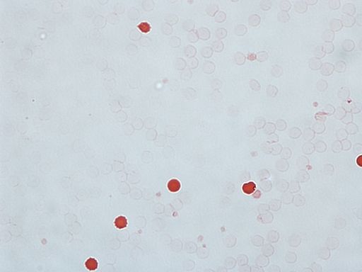
    Kleihauer Betke
    A KB stain positive for fetal blood, noted with the dark red cells.

Test Procedure:

  • Staining: A maternal blood sample is stained with a specific dye that highlights fetal red blood cells.
  • Microscopic Examination: The stained sample is examined under a microscope to calculate the percentage of fetal cells in the maternal blood.
  • Determining RhIg Dosage: The KB test results help determine the appropriate Rh immune globulin (RhIg) dose needed after a sensitizing event to prevent HDFN.

Associated Tests:

  • Fetal Bleed Screen: A preliminary, rapid test to detect if fetal and maternal blood have mixed. It identifies the presence of RhD positive red cells in an Rh-negative mother's blood.
  • Determining Further Action: A positive Fetal Bleed Screen typically leads to a KB test to decide if the standard RhIg dose suffices or if additional doses are required, considering one RhIg vial can protect against up to 30mL of RhD positive whole blood.

Significance for Rh-Negative Mothers:

  • The KB test, alongside the Fetal Bleed Screen, plays a vital role in managing Rh-negative pregnancies, ensuring the well-being of both mother and future pregnancies by accurately adjusting RhIg therapy to prevent HDFN.

COVID: Convalescent Plasma Therapy

What is Convalescent Plasma?

Convalescent plasma is a blood product that is obtained from individuals who have recovered from a particular infection, such as COVID-19. The plasma is collected through a process called plasmapheresis, which separates the plasma from the other components of the blood, such as red and white blood cells.

The plasma of recovered individuals contains antibodies that the body produced in response to the infection. These antibodies can potentially help fight the same infection in other individuals who are currently ill. Convalescent plasma therapy involves transfusing the plasma from recovered individuals into the bloodstream of patients who are currently suffering from the same infection, in the hope that the antibodies in the plasma will help boost their immune response and aid in their recovery.

COVID-19 Convalescent Plasma Therapy

 The COVID-19 pandemic has brought about unprecedented challenges to the healthcare industry, prompting researchers to look for innovative ways to fight the virus. One of the strategies that emerged was the use of convalescent plasma therapy, which involves using blood plasma from people who have recovered from COVID-19 to treat others who are still infected. Convalescent plasma therapy has been used in the past to treat other infectious diseases, including SARS and Ebola, but its effectiveness in treating COVID-19 was still under investigation.

The use of convalescent plasma as a treatment for COVID-19 was initially considered a promising approach, and it was authorized for emergency use in the United States by the Food and Drug Administration (FDA) in August 2020. However, subsequent studies and clinical trials have yielded mixed results on the effectiveness of convalescent plasma in treating COVID-19.

Convalescent plasma therapy served as a bridge between the early days of the pandemic, when no treatments were available, and the development of vaccines and monoclonal antibody therapy. When the pandemic first began, the world was not yet equipped with the tools and knowledge to fight the virus effectively. As a result, many people became severely ill or died, and hospitals were overwhelmed with patients. Convalescent plasma therapy provided an option to treat COVID-19 patients who were critically ill, with no other effective treatments available.

The use of convalescent plasma therapy was not without its challenges, however. The collection of plasma from recovered COVID-19 patients was difficult, and not all patients were eligible to receive the therapy due to various factors such as blood type compatibility and the timing of the illness. Additionally, the effectiveness of convalescent plasma therapy was not fully established, and the treatment was not widely available in all regions.

Some studies have suggested that convalescent plasma may have a modest benefit in reducing mortality rates and improving clinical outcomes in certain patient populations, such as those who receive plasma with high levels of antibodies early in the course of their illness. However, other studies have not found significant benefits from convalescent plasma therapy.

In February 2021, the National Institutes of Health (NIH) released updated guidelines on the use of convalescent plasma for COVID-19 treatment, stating that there was insufficient evidence to recommend either for or against its use. The guidelines noted that the available data were limited, and further research was needed to better understand the potential benefits and risks of convalescent plasma therapy.

Despite these challenges, the use of convalescent plasma therapy provided valuable insights into the treatment of COVID-19 and paved the way for the development of vaccines and monoclonal antibody therapy. Vaccines have proven to be the most effective way to prevent COVID-19, while monoclonal antibody therapy provides a targeted treatment for those who become infected with the virus.


How was the blood supply affected by COVID?

Blood Donation Decline Due To COVID 

Covid and blood
The COVID-19 pandemic has had a significant impact on blood donation worldwide. Blood donation centers and blood banks have seen a significant decline in the number of blood donations as a result of the pandemic, which has led to a shortage of blood products. In the early months of the pandemic, blood donation centers and blood drives were canceled or postponed due to lockdowns and social distancing guidelines, resulting in a sharp decline in blood donations. According to the American Red Cross, they experienced a significant decline in blood donations in March and April 2020, resulting in a shortage of nearly 200,000 blood products.

Reasons for Blood Donation Decline

Fear of COVID-19: One of the primary reasons for the decline in blood donations is the widespread fear of contracting COVID-19. Potential donors are apprehensive about visiting blood donation centers or participating in blood drives, concerned about virus exposure. In response, blood donation centers have implemented stringent safety measures, including:

  • Enhanced cleaning and disinfection
  • Social distancing protocols
  • Mandatory use of personal protective equipment (PPE), such as masks and gloves

Cancellation of Blood Drives: The enforcement of social distancing guidelines and lockdown measures has led to the cancellation of numerous blood drives and events. Schools, churches, and community centers, which are traditional venues for such drives, have faced closures or are operating at limited capacity, thereby hindering the organization of blood donation events.

Global Blood Shortages: The impact of the pandemic on blood donation has been global, with the World Health Organization (WHO) reporting shortages across many countries. These shortages pose significant risks for patients dependent on transfusions for life-saving treatments.

Continued Need for Blood Donations: Despite pandemic-related challenges, the demand for blood for surgeries, cancer treatments, and emergency care remains high. Blood donation centers are adopting additional safety protocols to protect donors and staff, urging healthy individuals to donate.

Efforts to Encourage Donation: To promote blood donation during the pandemic, centers have introduced measures such as:

  • Online registration and appointment scheduling
  • Incentives for donors, like gift cards or merchandise discounts

Reduced Demand and Supply Chain Disruptions: The postponement or cancellation of elective surgeries initially reduced the demand for blood. However, some areas saw increased demand due to COVID-19 related complications. Moreover, global travel restrictions and border closures disrupted the blood product supply chain, affecting availability in certain regions.

Operational Challenges: Additional safety measures required for blood collection and processing have impacted the efficiency of donation centers, resulting in:

  • Longer wait times for donors
  • Reduced blood collection capacity

Hospital Struggles with Blood Inventory: Hospitals faced severe shortages, resorting to splitting blood product units to extend supplies. Efforts were made to provide at least partial doses to those in need.

Loss of Long-Time Donors: Many regular donors have not returned to donate post-pandemic, leading to a decrease in the donor pool.

Ongoing Strains on Blood Supply: While there has been some recovery in the blood supply, challenges persist, particularly with the availability of O negative blood as of 2023.


Liver Issues and Transfusions

Liver veins
The Liver

How Can Liver Issues Cause Transfusion Need?

The liver plays a critical role in many important bodily functions, including detoxification, protein synthesis, and the production of bile. In certain medical situations, such as liver failure or drug overdose, the liver may become overwhelmed and unable to perform these functions adequately.

Liver issues can have a significant impact on the body's ability to clot blood, leading to an increased risk of bleeding. The liver is responsible for producing several clotting factors that are essential for the coagulation cascade, including factors II, V, VII, IX, X, and fibrinogen. In liver disease, the liver may not be able to produce enough of these clotting factors, leading to impaired blood clotting and an increased risk of bleeding, and thus a need for transfusion (potentially both plasma and Red Cells)

Patients with liver disease may experience bleeding from various sites, including the gastrointestinal tract, nose, gums, and skin. The severity of bleeding can vary, from minor bleeding to life-threatening hemorrhages. Additionally, liver disease can also lead to the development of portal hypertension, a condition where high blood pressure develops in the portal vein, leading to the formation of varices in the digestive tract that are prone to bleeding.

When patients with liver disease experience bleeding, they may require transfusions of blood products to replace the clotting factors that are deficient in their blood. Fresh frozen plasma (FFP) is a blood product that contains all of the clotting factors and is often used to replace deficient clotting factors in liver disease patients. In severe cases, patients may require transfusions of packed red blood cells to replace blood lost due to bleeding.

Esophageal Varices

Esophageal varices are swollen and fragile veins in the esophagus that can rupture and cause severe bleeding in patients with liver failure. Liver failure can result from various chronic liver diseases such as cirrhosis, hepatitis B or C, or alcohol abuse. When the liver is damaged, it can lead to an increased pressure in the veins that carry blood from the digestive organs to the liver (portal veins). This increased pressure can cause the veins in the esophagus to become enlarged and fragile, leading to the risk of bleeding.

Esophageal varices bleeding is a medical emergency and requires urgent medical attention. Patients may experience symptoms such as vomiting blood, black or tarry stools, low blood pressure, rapid heart rate, or confusion. Treatment typically involves stabilizing the patient's condition with fluids and blood transfusions, then endoscopic procedures such as band ligation or sclerotherapy to stop the bleeding and prevent future episodes.

In addition to treating the bleeding, it is important to address the underlying liver disease to prevent further damage to the liver and the development of additional varices. This may involve lifestyle changes such as avoiding alcohol and certain medications, as well as medical interventions such as antiviral therapies, immunosuppressants, or liver transplant in some cases.

Alcoholic Liver Disease

In advanced stages of alcoholic liver disease, the liver becomes severely damaged and is unable to perform its normal functions, including producing clotting factors that help the blood to clot, as mentioned This can lead to a condition called coagulopathy, where the blood is unable to clot properly.

Transfusions of blood or blood products may be necessary in patients with alcoholic liver disease who have coagulopathy to help prevent bleeding complications. For example, if a patient with alcoholic liver disease requires a surgical procedure or has a bleeding episode, transfusions of clotting factors or platelets may be needed to help stop the bleeding.

In addition, patients with alcoholic liver disease may develop anemia, which is a decrease in the number of red blood cells in the body. Anemia can cause fatigue, shortness of breath, and other symptoms. Transfusions of red blood cells may be necessary to help correct anemia in these patients.

It's important to note that while transfusions can be life-saving in some cases, they are not a cure for alcoholic liver disease. The underlying condition needs to be addressed through lifestyle changes, such as stopping alcohol consumption and improving nutrition, and in some cases, medical treatment may be necessary to manage complications of the disease.


Kidney Disease and Transfusions

Kidney disease is a serious health condition that affects millions of people worldwide. It is a condition that occurs when the kidneys are unable to function properly, resulting in a buildup of toxins and waste products in the blood. This can lead to various complications, including anemia, bleeding, and the need for transfusions.

The Kidneys Help Make Blood!

Why Would Patients With Kidney Disease Needs Transfusions?

Anemia: Kidneys play a vital role in producing a hormone called erythropoietin (EPO), which stimulates the bone marrow to produce red blood cells. In kidney failure, the kidneys may not produce enough EPO, resulting in anemia. Transfusions can be given to increase the number of red blood cells in the body and improve oxygen delivery to tissues.

Bleeding: People with kidney failure may be at a higher risk of bleeding due to the effects of uremia (accumulation of waste products in the blood), medications used to treat kidney failure, and other factors. In cases of severe bleeding, transfusions may be necessary to replace lost blood and prevent complications.

Uremia caused by Kidney Failure can contribute to bleeding in several ways:

  • Platelet dysfunction: Uremia can impair the function of platelets, which are small blood cells that play a crucial role in blood clotting. Platelets help to form a plug at the site of an injury and prevent excessive bleeding. In uremia, platelets may not function properly, leading to a higher risk of bleeding.

  • Vascular abnormalities: Uremia can cause changes in the walls of blood vessels, making them more fragile and prone to bleeding. These changes can include thickening, stiffening, and weakening of the vessel walls.

  • Coagulation abnormalities: Uremia can also affect the coagulation system, which is responsible for forming blood clots. In some cases, uremia can cause an imbalance in the coagulation system, leading to an increased risk of bleeding.

  • Medications: People with kidney disease may need to take medications to manage their condition, such as anticoagulants or antiplatelet drugs. These medications can increase the risk of bleeding, particularly if kidney function is impaired and the drugs are not cleared from the body efficiently.

Surgery: People with kidney failure may require surgery for various reasons, such as placement of a dialysis access or kidney transplant. Transfusions may be necessary during surgery to replace blood loss.

EPO Supplementation 

EPO supplementation can help increase the number of red blood cells in the body and improve oxygen delivery to tissues. EPO can be administered by injection under the skin or directly into a vein. The dosage and frequency of EPO supplementation may vary depending on the severity of anemia, the underlying cause of kidney failure, and other factors.

It is important to note that EPO supplementation can have potential risks and side effects, such as high blood pressure, blood clots, and an increased risk of certain types of cancer. Therefore, the decision to supplement with EPO should be made after careful consideration of the benefits and risks, and alternative treatments should be explored when possible.

In addition to EPO supplementation, other treatments for anemia in kidney failure patients may include iron supplements, vitamin B12 and folic acid supplements, blood transfusions, and medication adjustments to manage underlying conditions.


Massive Transfusion Protocol

Car Crash MTP
MTPs can be activated in severe trauma from a car accident
Massive transfusion protocol (MTP) is a critical medical intervention used in emergency situations to save the lives of patients with severe bleeding. This protocol is a set of guidelines developed by healthcare professionals that facilitate the rapid administration of large quantities of blood products, including red blood cells, plasma, and platelets. Studies have shown that early implementation of MTPs can improve patient survival rates and decrease the incidence of complications such as multiple organ failure, sepsis, and respiratory distress syndrome. By providing blood products in a targeted and timely manner, MTPs can help to replenish the patient's blood volume, restore normal clotting function, and improve tissue oxygenation.

We will discuss what MTP is, how it works, and why it is essential in emergency medical situations. We will also provide some insights on how MTP is initiated and what factors influence its success.

What is Massive Transfusion Protocol?

Massive Transfusion Protocol (MTP) is a pre-established set of procedures and guidelines for the administration of large volumes of blood products in trauma patients who have sustained significant blood loss. MTP is initiated in patients who have lost over 50% of their total blood volume, typically as a result of a severe injury or surgical procedure.

The goal of MTP is to rapidly replace the lost blood volume and maintain appropriate levels of oxygen delivery to the body's vital organs, including the brain, heart, and lungs.

How Does The Massive Transfusion Protocol Work?

MTP is a coordinated effort involving healthcare professionals from different specialties, including emergency medicine, critical care, hematology, transfusion medicine, and pharmacy. The protocol is typically initiated by the trauma team leader, who assesses the patient's condition and determines whether MTP is necessary.

Once the decision to activate MTP has been made, blood bank staff prepares and delivers a predetermined amount of blood products, including red blood cells, plasma, and platelets, to the patient's bedside. The blood products are then administered based on the patient's specific needs, as determined by laboratory tests and ongoing clinical assessments. MTPs are most successful when a 1:1 ratio of Red Blood Cells and Plasma is followed. So, for example, each MTP cooler may contain 6 RBCs and 6 units of plasma. Platelets and cryoprecipitate may also be prepared at specific intervals. 

Factors Influencing the Success of MTP

The success of MTP is influenced by several factors, including timely activation, appropriate selection of blood products, and effective monitoring of the patient's response to transfusion. The early initiation of MTP is critical, as it ensures that the patient receives blood products promptly, which can improve survival rates.

Another essential factor in the success of MTP is the selection of appropriate blood products. Red blood cells are typically administered first, followed by plasma and platelets in a predetermined ratio (usually 1:1). This approach ensures that the patient receives the necessary clotting factors to stop the bleeding.

Finally, effective monitoring of the patient's response to transfusion is essential. The patient's vital signs, laboratory values, and overall clinical condition must be continuously assessed to ensure that the transfusions are providing the intended benefit.

Why is MTP Important?

MTP is critical in emergency medical situations, as it can save the lives of patients with severe bleeding. Without MTP, patients who have lost significant amounts of blood may not receive the necessary blood products in a timely fashion, leading to poor outcomes and high mortality rates.

MTP has been shown to improve survival rates in trauma patients with severe bleeding, reducing the risk of death from bleeding by up to 70%. Additionally, MTP has been associated with a reduction in hospital length of stay and ICU admissions, leading to lower healthcare costs.

What are the negatives of activating an MTP?

While MTP can be life-saving, it is not without complications. Some potential complications of MTP include:

  • Transfusion reactions: Blood transfusions can trigger immune reactions in the recipient, leading to symptoms such as fever, chills, itching, hives, and difficulty breathing. These reactions can be mild to life-threatening.

  • Fluid overload: Rapid transfusion of large amounts of blood products can lead to fluid overload, which can cause edema, shortness of breath, and heart failure.

  • Coagulopathy: Patients who have lost a large amount of blood may also have impaired blood clotting function, which can be exacerbated by transfusions. This can lead to bleeding complications and make it difficult to control bleeding.

  • Hypothermia: Blood products that are stored at cold temperatures can cause hypothermia when rapidly infused into a patient. Hypothermia can cause a variety of complications, including cardiac arrhythmias and impaired coagulation function.

  • Transmission of infectious diseases: While blood products are tested for infectious diseases, there is still a risk of transmission of viruses, bacteria, or other pathogens.

  • Cost: MTP can be expensive due to the large amount of blood products used, which can strain healthcare budgets and resources.

  • Blood Wastage: Ocassionally, MTPs might be activated inappropriately, and thus blood products, especially thawed FFP, might be wasted. MTPs must only be called in true exsanguination emergencies. 

Ionized Calcium and Massive Transfusion

What is Ionized Calcium? Ionized calcium refers to the calcium in one's serum that isn't bound to proteins or other molecules. It plays a crucial role in various bodily functions, including blood clotting, heart function, and muscle contractions.

Challenges During Massive Transfusions: During massive blood transfusions, ionized calcium levels can drop due to several factors:

  • Dilutional Effect: Transfusing large volumes of blood products, which typically contain low levels of ionized calcium, can dilute the patient's ionized calcium concentration.

  • Citrate Anticoagulant: Blood products are preserved with citrate to prevent clotting. Citrate binds to calcium ions, reducing available ionized calcium. Although the liver metabolizes citrate, releasing calcium back into the blood, rapid transfusions can overwhelm this process.

  • Acidosis: Massive transfusions can increase blood acidity, leading to more calcium binding with proteins and less free ionized calcium.

Consequences of Ionized Calcium Depletion: A drop in ionized calcium during massive transfusions can lead to complications such as:

  • Impaired clotting function
  • Cardiac arrhythmias
  • Muscle cramps

Managing Calcium Levels: To counteract ionized calcium depletion, healthcare providers may administer calcium supplements like calcium gluconate or calcium chloride during and after the transfusion process. This intervention helps maintain essential physiological functions and prevent the complications associated with low ionized calcium levels.

This approach underscores the importance of monitoring and managing ionized calcium levels during massive blood transfusions to ensure patient safety and treatment efficacy.

How Do You Administer Blood Rapidly for an MTP?

The Belmont Rapid Infuser System: A Crucial Tool in Emergency Care

Overview of the Belmont Rapid Infuser: The Belmont Rapid Infuser is an advanced medical device designed for the rapid and efficient delivery of large volumes of fluids, including blood products, directly to patients. It's especially crucial in emergency situations where quick fluid replacement is essential.

How It Works:

  • Fluid Warming: The device can warm fluids to body temperature, ensuring patients receive fluids that match their physiological conditions.
  • Disposable Cassette System: Utilizes a unique cassette system with a built-in heating element and pump. Fluids, such as blood, are loaded into the cassette, warmed, and then infused at high speed.
  • Rapid Infusion Capabilities: Capable of infusing up to 1 liter of fluid in less than a minute, it's invaluable during massive transfusions required in trauma cases or surgical procedures.

Safety Features:

  • Air Embolism Prevention: Designed to minimize the risk of air embolism, a dangerous condition where air enters the bloodstream. It includes air detection sensors that halt infusion if air is detected, enhancing patient safety during rapid infusion.

The Role in Emergency Care: The Belmont Rapid Infuser is a vital component in the management of emergencies requiring fast fluid replacement. Its ability to quickly warm and infuse large volumes of fluids makes it a key device in trauma care and surgical settings, helping to save lives by stabilizing critical patients rapidly.